The blur caused by ocular aberrations is the main factor
limiting resolution
of images of the inside of the eye.
Adaptive optics is a technique used to compensate
for these aberrations. This work on adaptive optics has
come from David
Williams'
lab at the Center
for Visual Science, University of
Rochester where I spent 1996-1998
as a post-doc. To see the work done in Rochester,
follow
this link.
To see details of current AO research at the University of Houston, follow this link.
![]() Image of the retina before and after using adaptive optics
Image of the retina before and after using adaptive optics
 Average of many images of the same retinal location
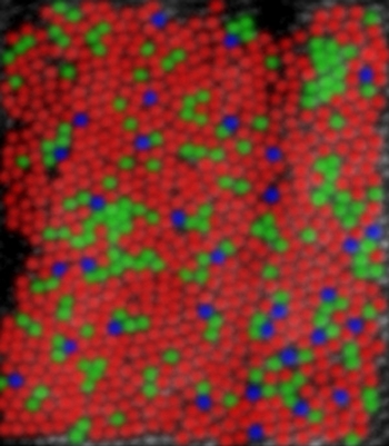
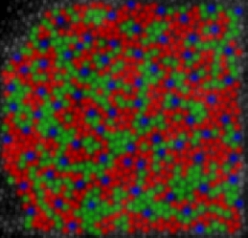
By combining retinal densitometry with the high resolution
images available using
adaptive optics, we were able to identify the short (S)-,
middle (M)-, and
long (L)-wavelength sensitive cones in the living human
eye. The following pictures
show examples from two subjects. In spite of the obvious
differences in
the proportion of L to M cones in these two subjects,
they both were found to have
normal color vision. You can read the details about how
we obtained these images in
our recent paper (Roorda, A. Williams, D.R. (1999) "The
arrangement of the three
cone classes in the living human eye", Nature397:
520-522).
 False-color image showing the arrangement of cones for subject JW
at a location 1 deg nasal from the central fovea
 False-color image showing the arrangement of cones for subject JW
at a location 1 deg temporal from the central fovea
 False-color image showing the arrangement of cones for subject AN
at a location 1 deg nasal from the central fovea
 False
color image showing the arrangement of cones in a macaque
False
color image showing the arrangement of cones in a macaque
monkey at a location 1.4 deg nasal from the central fovea
SML locations in an EXCEL spreadsheet.
SML locations in an HTML document (Internet Explorer only).
An analysis of the packing arrangement of these cone arrays
is published in Vision
Research.
Roorda, A., Metha, A., Lennie, P., Williams, D.R., “Packing
Arrangement of the Three
Cone Classes in the Primate Retina” Vision Research,
41(12) 1291-1306 (2001).
last updated: Feb 17, 2003