The overarching question that motivates the research in the Roorda lab is: How do humans convert the two-dimensional images that land on the retina into such a rich perceptual experience?
Specifically....
- What limits do the imperfections in the optical system of the eye impose for human vision?
The left panel shows a typical human PSF (6 mm pupil),
the center frame shows a 20/20 sized E,
the third frame shows how that E would appear on the retina.
- How do we acheive acuity levels near the photoreceptor sampling limit with ever-present fixational eye movements?

AOSLO video of human retina viewing
a letter 'E' which is projected onto its surface.
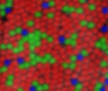
- How do we convert 2-D images sampled by a random arrangement of three cone types into our exquisite sensitivity to color and 3D space?
Image showing the distribution of the three
classes of human cone photoreceptor,
sensitiive to bluish, greenish and reddish
regions of the visible light specturn.
To tackle these questions, members of the lab design and build optical instruments that (i) correct the eye's imperfections (ii) track the retina with high speed and accuracy and (iii) measure visual structure and function on a cellular scale.
Our favorite tool is
adaptive optics - a technology
originally developed for astronomical imaging from ground-based telescopes - to
correct the eye’s aberrations and to image and/or present stimuli to the retina
with unprecedented resolution. Overcoming optical limitations with adaptive
optics has enabled new discoveries in vision science, from mapping
the trichromatic cone mosaic to revealing how human
visual acuity improves with aberration correction. See our
publication page for more details.